Properties (17)
- purpleRed
- (Ajmera et al., 2026)
•
Altered intrinsic brain connectivity in misophonia, with and without hyperacusis
Status:: #status/reviewed
Metadata
- Item Type: #journalArticle
- Authors:: Shagun Ajmera, Rafay A. Khan, Gibbeum Kim, Namitha Jain, Ariana Castro, Howard Berenbaum, Fatima T. Husain,
- Date:
- Date Added: 1769137375000
- URL: https://linkinghub.elsevier.com/retrieve/pii/S0378595525003387
- DOI: 10.1016/j.heares.2025.109521
- Cite Key: ajmeraAlteredIntrinsicBrain2026
- Topics: [object Object]
- Tags:: #reviewed, #Misophonia, #Neurology, #seminal-work, #in-review, #hyperacusis, #machine-learning, #Fmri, #sound-tolerance-disorders, #resting-state-connectivity,
- PDF Attachments
Abstract
| Background/Purpose | Background- • Decreased Sound Tolerance disorders (DST) are characterized by reduced endurance or heightened sensitivity to auditory stimuli provoking adverse emotional or physiological reactions. • Misophonia and loudness hyperacusis frequently co-occur; more severe misophonia symptoms correlate with hyperacusis presence. • Prior neuroimaging studies of misophonia reported heightened cortical and subcortical activations to trigger sounds in salience network and auditory cortex. • Altered brain interactions during trigger sound exposure involve emotion regulation networks and subcortical emotion processing regions. • Recent findings suggest significant motor cortex role in misophonia, possibly reflecting orofacial hyper-mirroring tendencies. • Previous studies lacked data-driven whole-brain approaches and relied on seed-based analyses with predefined regions. • Most studies lacked structured psychiatric assessments for misophonia validation, relying on self-report questionnaires. • Control groups typically did not screen for hyperacusis, potentially confounding misophonia-specific neural patterns. Purpose- • Identify neural connectivity-based markers of misophonia among cortical and subcortical networks using resting-state fMRI. • Evaluate detectability of misophonia in isolation (MI) and when comorbid with hyperacusis (MH) using machine learning. • Address gaps by categorizing participants into misophonia-only and misophonia-with-hyperacusis groups based on structured psychological evaluations. • Utilize data-centric machine learning on whole-brain interactions with rigorous model training. • Dissociate connectivity trends affiliated with misophonia in the comorbid group. |
| Participants/samples | • N = 76 total; misophonia-only (MI) n = 27; misophonia & hyperacusis (MH) n = 24; controls (CTR) n = 25. • University students aged 18–25 years. • MI group: age 21 ± 2, 20F/7M; MH group: age 20 ± 2, 18F/6M; CTR group: age 20 ± 2, 13F/12M. • No group differences in age, sex, or pure tone audiometry hearing thresholds. • Inclusion based on structured clinical interviews using Amsterdam Misophonia Scale (A-MISO-S) and diagnostic criteria from Lewin et al. (2015). • MH group had significantly lower loudness discomfort levels (LDLs) compared to CTR. • All participants had no tinnitus. |
| Measures | • Misophonia Questionnaire (MQ; Wu et al., 2014) – self-report measure of misophonia symptoms and severity. • Hyperacusis Questionnaire (HQ; Khalfa et al., 2002) – self-report assessment of hyperacusis presence and severity. • Amsterdam Misophonia Scale (A-MISO-S; Schröder et al., 2013) – semi-structured interview and clinical rating scale for misophonia symptom severity. • Loudness Discomfort Levels (LDLs) – audiological assessment measuring tolerance for pure tones (0.5 kHz, 4 kHz) and speech stimuli. • Structured Clinical Interview for DSM-5 – assessment of major depressive disorder symptoms. • Resting-state fMRI functional connectivity – Pearson correlation between BOLD time series of 414 brain regions (400 cortical ROIs from Schaefer atlas, 14 subcortical ROIs from Harvard-Oxford atlas). |
| Procedures | • Resting-state fMRI (10 min; eyes open fixation). • Whole-brain parcellation (400 cortical + 14 subcortical ROIs). • 85,491 functional connections extracted per participant. • Elastic-net logistic regression classifiers (MI vs CTR; MH vs CTR). • Cross-validation and hyperparameter optimization. • Network-level aggregation of connectivity. |
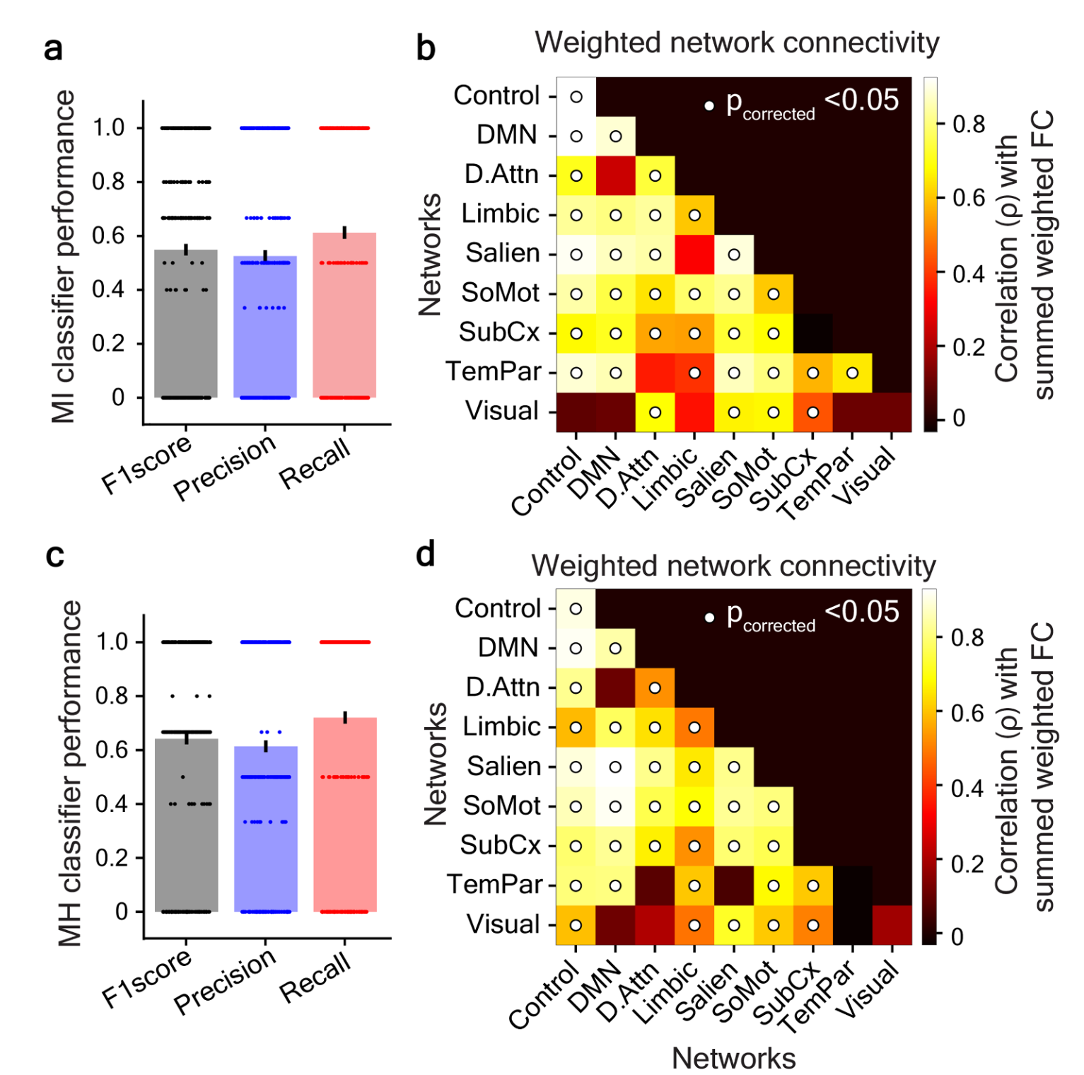
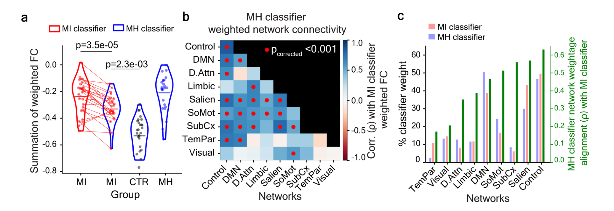
| Results | MI vs. CTR classification- • MI classifier distinguished MI from CTR. • Consistently highly weighted FC edges concentrated in specific brain regions: decreased connectivity in MI for globus pallidus, posterior middle temporal gyrus, anterior insula, right lateral prefrontal cortex, posterior cingulate cortex; increased connectivity for inferior parietal lobule. • Network-level analysis revealed significant connectivity alterations in salience, somatomotor, and frontoparietal control networks. • Salience network connectivity with all brain networks showed high correlation with full FC model. • Somatomotor network connectivity with all brain networks showed high correlation with full FC model. • Frontoparietal control network connectivity significant for all networks except visual network. MH vs. CTR classification- • MH classifier distinguished MH from CTR. • Consistently highly weighted FC edges concentrated at anterior medial PFC, dorsolateral PFC, primary motor and supplementary motor areas. • Network-level analysis revealed alterations in salience, somatomotor, and frontoparietal control networks similar to MI classification. • Additional unique alterations in visual network connectivity with control and limbic networks specific to MH group. Overlap and specificity- • MH classifier distinguished MI from CTR with significant separation. • Individual ordering of MI group preserved across MI and MH classifier weights. • 33 of 36 significant network pairs in MH classification also significant in MI classification. • Network pairs unique to MH classification involved visual network with control and limbic networks. • Misophonia-aligned network pairs in MH identification carried 76.25% of total MH classifier weight. Subcortical connectivity- • MI classifier assigned non-zero weights to hippocampus, globus pallidus, and striatal regions (putamen, accumbens, caudate). • MH classifier assigned zero weights to all subcortical connections. • Net weighted subcortical intraconnectivity significantly different between MH and MI groups. • No significant difference in subcortical connectivity between MH and CTR groups. |
| Discussion/Limits | Conclusion- • The authors conclude that machine learning identified distinct whole-brain functional connectivity patterns that reliably distinguish misophonia from controls. • The findings indicate that core misophonia-related disruptions consistently involve salience, somatomotor, and frontoparietal control networks, reflecting impairments in emotion regulation, motor inhibition, and attentional control. • The data support that cognitive control deficits represent a central and previously underappreciated feature of misophonia. • The findings indicate that misophonia and hyperacusis have partially dissociable neural mechanisms, with core misophonia-related patterns persisting across both groups while hyperacusis contributes additional alterations in visual and higher-order networks. • The authors conclude that subcortical connectivity alterations, particularly in basal ganglia (e.g., globus pallidus), are specific to misophonia-only, may reflect motor simulation processes, and suggest overlap with OCD-related neural mechanisms. • The findings indicate that hyperacusis co-occurrence can obscure misophonia-specific neural signatures, emphasizing the need for careful diagnostic separation. • The data support that distinct neural patterns necessitate targeted, psychology-based interventions, with cognitive-behavioral approaches aimed at emotion regulation, cognitive reappraisal, and attentional control as promising treatments. |
| Limitations:: | • Conclusions about active role of basal ganglia in trigger-sound processing based on resting-state data require validation with task-based neuroimaging. • Whether globus pallidus plays similar role in perception of non-orofacial human-generated sounds remains unknown. • Resting-state brain connectivity may be more apparent in MI than MH; larger samples of task-based neuroimaging may reveal OCD-associated differences in MH group. • Research into neural bases of misophonia and hyperacusis is in early stages. • Behavioral aspects involving emotions like disgust and aversive learning not fully integrated with neuroimaging findings. |
| Future Directions:: | • Task-based neuroimaging needed to test active role of basal ganglia in trigger-sound processing. • Investigation needed whether globus pallidus role extends to non-orofacial human-generated sounds. • Larger task-based neuroimaging samples needed to reveal potential OCD-associated differences in MH group. • Research should focus on translating insights into clinical interventions grounded in neurological and psychological characteristics specific to misophonia. • Development of standardized CBT protocols tailored to modulate dysregulated activity in identified neural networks. |
| Relevance:: | • A BIG POINT her is that there was no task of stimulus exposure. it was purely recording passive resting state fMRI, then using machine learning to classify subjects based on connectivity data. • used exploratory approach to look at connectivity of people with Misophonia with/without hyperacusis and full controls. training a classifier model on fMRI data, they got models that correctly distinguished the groups 63-67% of the time • found a few abnormal connectives specific to each group • a good source for using machine learning /classifier models to distinguish Misophonia neural data from controls • results show more refined neurobiological distinction between Misophonia and hyperacusis. • results support the Savard and Coffey 2025 cognitive model of Misophonia • results support the (Kumar et al., 2021) motor model of Misophonia • provides data supporting the use of CBT and other psychology-based therapeutic approaches to Misophonia |
Notes
key terms and concepts
- misophonia – debilitating sound intolerance condition marked by extreme emotional and physiological responses to everyday sounds, particularly human-generated sounds (e.g., chewing, slurping).
- loudness hyperacusis – decreased sound tolerance disorder involving aversive response to everyday sounds perceived as abnormally loud.
- resting-state functional connectivity (FC) – measure of spontaneous neural interactions between brain regions during rest, assessed via correlation of fMRI BOLD signal time series.
- machine learning classification – data-driven multivariate approach using elastic-net regularized logistic regression to distinguish groups based on functional connectivity patterns.
- salience network – functional brain network implicated in emotion processing and detection of behaviorally relevant stimuli.
- frontoparietal control network – brain network involved in cognitive control, attentional regulation, and motor inhibition.
- somatomotor network – brain network associated with motor execution and sensorimotor processing.
- subcortex – deep brain structures including basal ganglia (striatum, globus pallidus), hippocampus, amygdala, and thalamus.
my engagement notes
my questions
my comments
Bookmarks
-
bookmark talking about the Subcortical alterations in misophonia-only: links to motor simulation and OCD-like traits (page 8)
-
bookmark for talking about OC trait connection (page 8)
reading notes
contextual statement
In a 2026 study, Ajmera attempted to identify neural connectivity-based markers of misophonia using an optimized machine learning framework applied to resting-state fMRI data from 76 university students (ages 18–25) categorized into misophonia-only, misophonia-with-hyperacusis, and control groups based on structured clinical interviews and established questionnaires.
summary statement
In a 2026 study, Ajmera aimed to identify neural connectivity markers distinguishing misophonia in isolation and when comorbid with hyperacusis by training machine learning classifiers on resting-state fMRI functional connectivity from 76 young adults rigorously categorized via structured interviews. They found that both misophonia-only and comorbid groups showed altered connectivity in salience, somatomotor, and frontoparietal control networks, with misophonia-only specifically showing subcortical basal ganglia disruptions and the comorbid group showing unique visual network alterations. From this they concluded that core misophonia-related brain alterations persist despite hyperacusis comorbidity, that subcortical connectivity dissociates the two conditions, and that the findings support psychology-based (CBT) therapy targeting dysfunctional connectivity in salience and control circuits for treating misophonia.
zettlekasten
calls for future work on task based neuroimaging and informing future interventions
machine learning approach top Misophonia classification
support for psychology-based therapeutic approaches (CBT) to Misophonia
connectivity data supports the Kumar motor basis model of Misophonia
neural connectivity findings supporting the Savard and Coffey 2025 cognitive model of Misophonia