Properties (15)
- purpleRed
- Multichannel auditory cortical responses in misophonia
- (Karupaiah et al., 2025)
•
Multichannel auditory cortical responses in misophonia: A neurophysiological investigation
Status:: #status/reviewed
Metadata
- Item Type: #journalArticle
- Authors:: Kamalakannan Karupaiah, Rakesh Trinesh, Ajith Kumar Uppunda, Prashanth Prabhu,
- Date:
- Date Added: 1769374192000
- URL: https://linkinghub.elsevier.com/retrieve/pii/S037859552500276X
- DOI: 10.1016/j.heares.2025.109458
- Cite Key: karupaiahMultichannelAuditoryCortical2025
- Topics: [object Object]
- Tags:: #Misophonia, #reviewed, #EEG, #ALLR, #auditory-long-latency-resposne, #cortical-auditory-processing,
- PDF Attachments
Abstract
| Background/Purpose | Background- • Misophonia defined by intense emotional/physiological responses to specific auditory triggers; neurophysiological basis remains unclear. • fMRI studies show heightened activation in amygdala, anterior insula, auditory cortex, and ACC in misophonia. • Prior EEG work (Schröder et al., 2014) reported reduced N1 amplitude using pure tones; proposed as neurophysiological biomarker. • MMN reductions also documented, suggesting altered pre-attentive processing (Schröder et al., 2013; Patro et al., 2025). • Aryal & Prabhu (2023) found earlier P1/N1 latencies and reduced P1-N1 amplitudes using tone burst stimuli. • Multichannel EEG provides spatial distribution of neural generators with high temporal resolution, enabling topographic analysis not available in single-channel paradigms. • Prior CAEP studies did not characterize scalp-wide spatiotemporal distribution; no multichannel ALLR study in misophonia existed. Purpose- • Investigate multichannel ALLR (P1-N1-P2) in individuals with and without misophonia using a non-trigger speech stimulus. • Compare peak latencies and amplitudes at midline sites (Fz, Cz, Pz). • Characterize scalp-wide spatiotemporal distribution differences between groups. • Identify potential electrophysiological markers for diagnosis and treatment monitoring. |
| Participants/samples | • N = 30 female participants; ages 18–30. • Misophonia group: n = 15; MisoQuest ≥61, RAMISO-S ≥11 (mild–moderate severity range). • Control group: n = 15; MisoQuest ≤16, RAMISO-S 0–10 (subclinical). • All-female sample; not a priori restriction, outcome of recruitment. • Exclusion: tinnitus, hyperacusis, phonophobia, hearing loss, otological disorders, ototoxic medication, noise exposure. • Normal peripheral hearing confirmed via PTA, tympanometry, DPOAEs. |
| Measures | • MisoQuest (presence of misophonia) • RAMISO-S (severity of misophonia) • Tinnitus Handicap Inventory, Modified Khalfa Hyperacusis Questionnaire, DSTS-S (exclusion screening) • 32-channel EEG (Quik-Cap Neo Net, Grael amplifier; Compumedics); 10–20 system; referenced to right mastoid • ALLR peak latency and amplitude at Fz, Cz, Pz for P1, N1, P2 • Scalp topography via cluster-based permutation testing (BESA Statistics v2.1) |
| Procedures | • Passive listening paradigm; participants watched silent closed-captioned video to maintain alertness. • Stimulus: voiced alveolar stop /da/, 130 ms duration, 80 dB SPL, ISI 2000 ms; 200 sweeps. • Stimulus confirmed not to overlap with any participant's personal misophonic triggers. • EEG: 0.1–100 Hz online bandpass, 1024 Hz sampling; offline processed in BESA (0.1 HP, 30 Hz LP). • Artifact rejection: ocular correction via BESA algorithm; epoch threshold ±100 µV; spherical spline interpolation for ≤2 bad channels. • Waveform analysis: M1-M2 average reference; spatiotemporal analysis: average reference. • Epochs: −100 to 500 ms; peak identification automated then manually verified by two experts. • Statistics: independent samples t-tests (two-tailed); Bonferroni-Holm correction for amplitude comparisons; Shapiro-Wilk normality confirmed. |
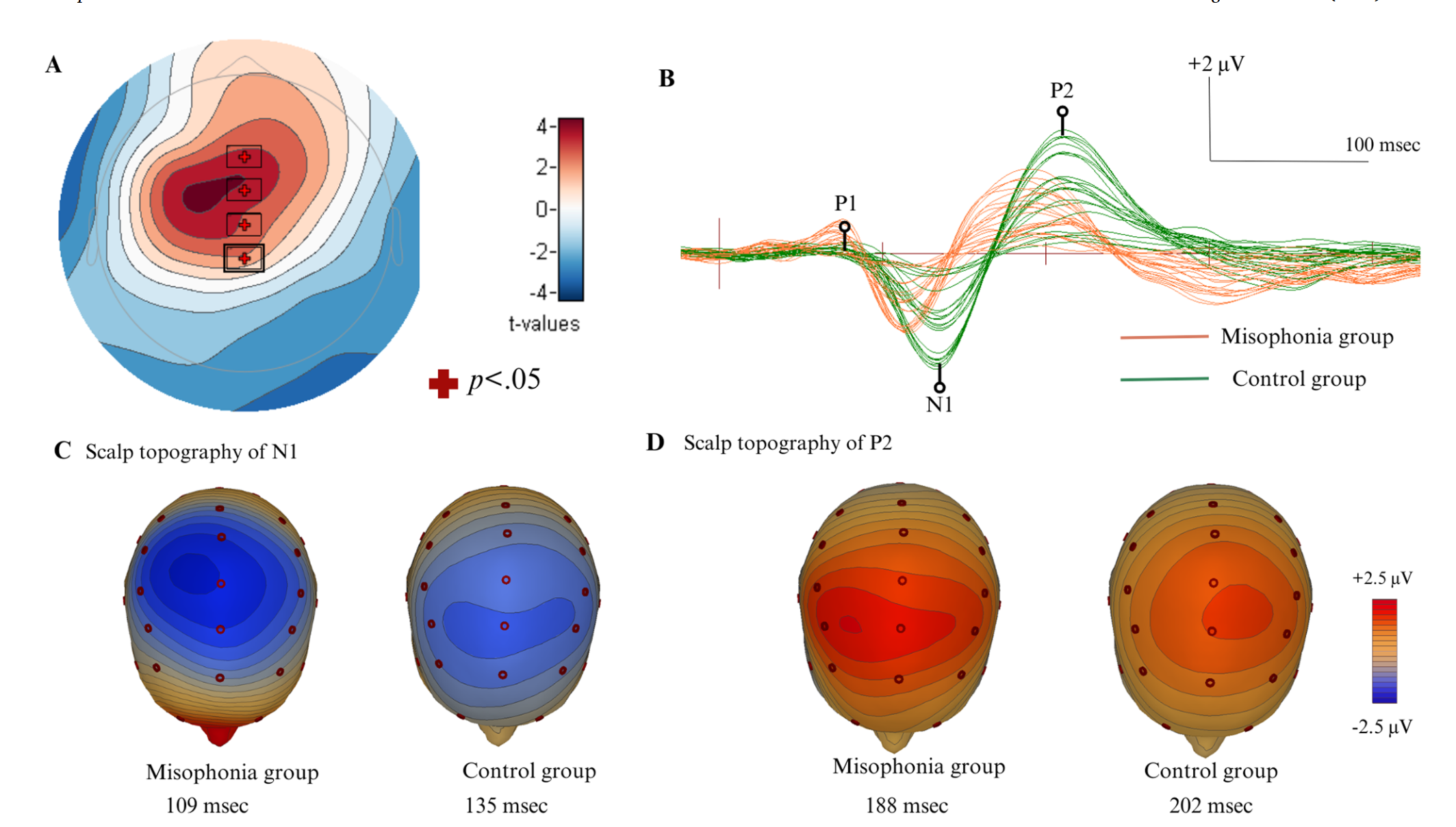
| Results | • Misophonia group showed earlier latencies across all P1-N1-P2 peaks at all three electrode sites (Fz, Cz, Pz); large effect sizes throughout. • N1 amplitude was significantly reduced in the misophonia group at all three electrode sites; P1 and P2 amplitudes did not differ significantly between groups. • Scalp topography differed significantly between groups in a ~198–254 ms window, primarily over Fz, FPz, Cz, CPz. • At N1 peak: misophonia group showed centro-parietal distribution; controls showed fronto-central distribution. • At P2 peak: misophonia group showed frontal distribution; controls showed central distribution. • Butterfly plots confirmed earlier latency and reduced N1 amplitude in misophonia grand average waveforms. |
| Discussion/Limits | Conclusion- • The authors conclude that earlier P1-N1-P2 latencies indicate faster neural transmission or heightened neuronal excitability at generators of the P1-N1-P2 complex (superior temporal gyrus, primary auditory cortex, subcortical regions). • The findings indicate earlier latencies may reflect overactive auditory-limbic pathways consistent with anterior insula hyperactivity documented in neuroimaging studies. • The authors conclude it remains equally plausible that accelerated processing is a consequence of misophonia rather than a predisposing cause, consistent with neuroplastic changes from repeated emotional reactivity. • The data support reduced N1 amplitude as a candidate neurophysiological biomarker of misophonia, replicating Schröder et al. (2014) with a speech stimulus. • The findings indicate altered spatial distribution of auditory cortical responses (centro-parietal vs. fronto-central), suggesting atypical spatial organization of auditory processing. • The authors conclude the observed patterns may reflect auditory-limbic dysregulation consistent with anterior insula and emotion-network hyperactivity. • The findings indicate that using non-trigger stimuli reveals fundamental cortical processing differences independent of emotional reactivity. |
| Limitations:: | • All-female sample limits generalizability. • No formal diagnostic criteria for misophonia; group assignment based solely on questionnaire cutoffs. • Non-trigger stimulus (/da/) precludes conclusions about trigger-specific vs. generalized auditory differences. • Small sample (n = 15 per group). • No severity subgroup analysis (all misophonia participants fell in mild–moderate range). • Cannot determine whether electrophysiological differences are predisposing or acquired. |
| Future Directions:: | • Replicate with trigger-specific stimuli (chewing, swallowing, lip-smacking) to test generalizability of N1 findings. • Longitudinal and interventional studies examining whether ALLR markers change with treatment. • Studies across misophonia severity levels. • Source localization and functional connectivity analyses to map cortical/subcortical networks. • Evaluate clinical utility of multichannel ALLR for diagnosis and treatment monitoring. |
| Relevance:: | • looked at differences in auditory late latency responses (ALLR) resposnes using 32 channel EEG , focusing on P2-N1-P2 peaks in Misophonics vs controls using a voiced consonant as a stimulus • findings showed earlier and reduced N1 amplitudes in the Misophonia group • results conflict with (Schröder et al., 2014) who didn’t find any differences in N1 latency, though this may be due to methods differences • also found the Misophonia group to show a centro-parietal activation topography compared to the control groups fronto-central • proposes reduced N1 amplitude as a possible biomarker for Misophonia |
Notes
key terms and concepts
Auditory Late Latency Response (ALLR) – cortical auditory evoked potential comprising P1-N1-P2 peaks reflecting early cortical auditory processing and attentional mechanisms; recorded ~50–250 ms post-stimulus onset.
N1 component – prominent negative peak within ALLR associated with attention allocation to auditory stimuli; proposed neurophysiological biomarker of misophonia.
Scalp topography – spatial distribution of electrical potentials across the scalp reflecting underlying neural generator patterns; analyzed via cluster-based permutation testing.
Sensory gating – neural inhibitory mechanism reducing cortical responses to redundant or irrelevant stimuli; impairment implicated in misophonic hyperreactivity.
Cluster-based permutation testing – nonparametric statistical method controlling family-wise error rate across electrode-time comparisons; 2000 permutations used.
MisoQuest – 14-item self-report questionnaire for presence of misophonia; scores ≥61 indicate misophonia.
RAMISO-S – Revised Amsterdam Misophonia Scale; quantifies misophonia severity (0–10 subclinical; 11–20 mild; 21–30 moderate; 31–40 severe).
my engagement notes
my questions
my comments
Bookmarks
reading notes
contextual statement
In a 2025 study, Karupaiah et al. investigated multichannel auditory late latency responses (ALLR) in misophonia by comparing P1-N1-P2 peak latencies, amplitudes, and scalp topographies between 15 misophonic and 15 control adult females using a 32-channel EEG passive listening paradigm with a non-trigger speech stimulus (/da/).
summary statement
In a 2025 study, Karupaiah et al. examined auditory cortical processing in misophonia by recording multichannel ALLR to a non-trigger speech syllable in matched misophonia and control groups. They found that individuals with misophonia showed earlier latencies across all P1-N1-P2 peaks, selectively reduced N1 amplitudes, and distinct centro-parietal (vs. fronto-central) scalp topographies compared to controls. From this they concluded that misophonia involves generalized alterations in early auditory cortical processing and spatial generator organization, with reduced N1 amplitude proposed as a candidate neurophysiological biomarker, though whether these differences are predisposing or acquired remains unresolved.
Zettelkasten
contrasting findings from Schroder 2014
Idea of observed processing differences being a result of rather then cause of Misophonia
topographical and spatiotemporal differences in Misophonia