Properties (1)
- the larger context of this major topic is in the linked parent note
- this note is to hold all data points (observations) implications/theoretical ponderings about the role of/impact on physiological/neurophysiological factors in Misophonia
- attempts should be made to provide links to relevant images in the Misophonia neurophysiology Atlas excalidraw file
- see the outline Guide for more details on placement/segmentation of notes
- a note belongs here if it meets one of the following
- describes abnormalities in physiological (ex: heart rate variability, GSR, pupillometry) in Misophonics compared to controls. this can be specifically in response to some stimulus presentation, task, or jsut a general systemic difference between Misophonics and controls.
- describes abnormalities in neurophysiology (ex: fMRI structures or connectivity, BOLD responses, EEG) in Misophonics compared to controls. this can be specifically in response to some stimulus presentation, task, or jsut a general systemic difference between Misophonics and controls.
- a note does NOT belong here if it meets one of the following
- describes a neuro/physiological model of Misophonia. if a point supports/is used to support a model, make a brief mention of the model with a block link to the model notes page.
- neurophysiological
- physiological
1. Neural Network Mechanisms
Misc.
bold-headers
the influence of context on the emotional responses to sounds in and out side of Misophonia suggests the involvement of the ventral auditory processing stream
in their 2025 literature review, Savard et all note that the ventral auditory processing stream is involved in making perceptual decisions regarding source identification and recognition ( see the process known as “auditory-scene analysis”) (Savard & Coffey., 2025)
bold-headers
connectivity data supports link to OCD like traits in Misophonia and a possible differentiator for other sound issues
In a 2026 study, Ajmera used an optimized machine learning framework applied to resting-state fMRI data to identify neural connectivity-based markers of misophonia across individuals categorized as misophonia-only, misophonia with hyperacusis, and controls. The findings indicate that misophonia exhibits OCD-related traits at a neural level, particularly through altered connectivity in basal ganglia and frontoparietal control circuits, suggesting shared mechanisms involving compulsivity, motor inhibition, and intolerance-related processes; importantly, because these subcortical alterations were observed specifically in the misophonia-only group and not in the comorbid group, the results imply that OCD-like neural features may be more central to “pure” misophonia rather than reflecting general sound intolerance or hyperacusis (Ajmera et al., 2026).
bold-headers
machine learning approach top Misophonia classification
a 2026 study used a data-driven machine learning approach in which elastic-net regularized logistic regression was applied to whole-brain resting-state functional connectivity data to identify a sparse subset of informative connections that could reliably distinguish misophonia groups from controls, while cross-validation ensured generalizability and the resulting model weights provided interpretable neural markers of the condition. The findings showed that classification was driven by consistent alterations in salience, somatomotor, and frontoparietal control networks, with additional subcortical disruptions specific to misophonia-only. These results imply that machine learning can not only detect misophonia from brain data but also isolate clinically meaningful neural circuits that can guide targeted, mechanism-based interventions (Ajmera et al., 2026).
bold-headers
Visual-Auditory Discomfort Overlap and Cross-Modal Cortical Hyperexcitability in Misophonia
In their 2025 study, Haigh examined whether visual and auditory discomfort overlap and whether auditory discomfort reflects cortical hyper-responsivity. An online survey showed that pattern glare was significantly associated with hyperacusis and functional hearing more strongly than misophonia, and a NIRS study found that an uncomfortable 16 Hz tone elicited larger and slower oxyhemoglobin responses than a 2 Hz tone, with no significant deoxyhemoglobin amplitude differences. The authors concluded that visually sensitive individuals are more likely to report pain-related auditory sensitivity, supporting a shared hyperexcitable cortical mechanism across modalities. They further suggest that pain-focused and emotion-focused sensitivities may form distinct clusters and that peak amplitude and latency reflect separable components of auditory discomfort. (Haigh et al., 2025)
bold-headers
Surja findings of normal audiological functioning in Misophonia
In a 2023 study, Suraj assessed linear OHC, non-linear OHC, and MOCB efferent functioning in misophonia by comparing TEOAE amplitudes, DPOAE amplitudes, and contralateral suppression magnitudes between misophonic and non-misophonic adults with normal hearing. They found no group differences in absolute OAE amplitudes or in suppression magnitudes for either TEOAEs or DPOAEs at any frequency. From this they concluded that cochlear OHC mechanisms and MOCB efferent functioning are not major contributors to misophonia, and that the pathophysiology likely lies more centrally within the auditory and limbic systems (Suraj et al., 2023).
bold-headers
Insula-Sensorimotor Connectivity Beyond the Mirror-Neuron Account: Neural Evidence for Non-Orofacial Triggers
in there 2022 fMRI study, Hansen challenge the “motor basis” account of misophonia proposed by Kumar et al. (2021) by demonstrating that altered connectivity in misophonia is not specific to ventral premotor cortex (PMv), the region typically associated with mirror neurons. Instead, they found increased insula connectivity across multiple sensorimotor regions, including primary motor (M1), primary somatosensory (S1), and dorsal premotor cortex (PMd), suggesting that the effect is not confined to a mirror-neuron mechanism. Moreover, by functionally defining finger-related motor and somatosensory regions, they showed that individuals with higher misophonia scores also exhibit differential insula connectivity with finger representations, providing neural evidence for non-orofacial triggers. This broader pattern of sensorimotor–insula connectivity undermines the claim that misophonia is primarily driven by orofacial mirroring and instead supports a more distributed neural account that includes direct representation of non-oral triggers rather than their acquisition solely through associative learning. (Hansen et al., 2022)
-
bold-headers
Hansen 2026 on the need to look beyond orofacial motor areas in Misophonia
in a 2026 fMRI study, Hansen discusses how Recent studies show increased anterior insula connectivity with the supplementary motor cortex and precentral gyrus in misophonia. This has been interpreted through an orofacial action perception and mimicry framework. Hansen goes on to argue that findings of co-occurring misokinesia (movement aversion) may also contribute to motor cortex involvement. in the Hansen 2026 study, they Notably found peak connectivity in the superior/medial motor cortex and extension into the postcentral (somatosensory) gyrus, suggesting that mechanisms beyond orofacial and purely motor explanations should be considered. (Hansen et al., 2026)
- see narrative contrast note for large ready made blurb on this topic
bold-headers
White Matter Structural Differences Supporting Misophonia as a Distinct Neurobiological Condition
In a 2021 study, Eijsker investigated whether Misophonics exhibit white matter structural abnormalities by comparing fMRI data for 24 Misophonics and 25 healthy matched controls. Misophonics showed greater white matter volume and denser nerve fibers in three key pathways: the inferior fronto-occipital fasciculus, the anterior thalamic radiation, and the body of the corpus callosum. These abnormalities were concentrated in circuits governing social-emotional processing and the automatic capture of attention by aversive or threatening stimuli, both of which are centrally relevant to the misophonic experience. The authors concluded that these structural differences provide meaningful neurobiological evidence in support of misophonia as a distinct clinical condition with a measurable basis in brain architecture (Eijsker et al., 2021).
bold-headers
affective switching deficits in Misophonia related to function of OFC
Black et al. (2026) link the observed affective switching deficits in misophonia to the role of the orbitofrontal cortex (OFC), noting that effective inhibition is essential for set-switching by suppressing competing task sets and minimizing interference during transitions. In misophonia, other works have shown the OFC shows hyperactivation specifically in response to trigger stimuli, and the authors argue this heightened activity reflects the OFC's role in facilitating functional synchronization across brain regions in ways that may directly influence behavioral task performance. From this they conclude that the reduced affective flexibility observed in misophonia is not simply a cognitive phenomenon but is grounded in identifiable neural dysregulation, with OFC hyperactivation serving as a plausible neural mechanism linking misophonic reactivity to impaired inhibitory control during affective switching (Black et al., 2026) .
bold-headers
speculative role of medial superior olive (MSO)
in a 2026 study, Roy et al found normal ILD thresholds alongside elevated ITD thresholds in Misophonics vs controls. the authors suggest that this suggests functional deficits in brainstem circuits responsible for binaural temporal integration. they further point to the medial superior olive (MSO) due to its role in processing fine timing cues needed for spatial hearing, but they note that this interpretation is speculative and requires direct confirmation. (Roy et al., 2026)
bold-headers
background info on the anterior insular cortex in the context of Misophonia
review via (Hansen et al., 2026)
- the anterior insular cortex is implicated in a number of Misophonia relevant functions, including disgust, subjective pain evaluation, goal directed attentional control, and interoception (Hansen et al., 2026)
- Early fMRI studies consistently show that individuals with misophonia exhibit heightened activation to trigger stimuli in the anterior insula, often alongside increased activation in related regions such as the anterior cingulate cortex, hippocampus, and ventromedial prefrontal cortex, compared to neurotypical controls. (Kumar et al., 2017), (Schröder et al., 2019), (Grossini et al., 2022)
- Functional connectivity studies indicate that individuals with misophonia show stronger insula coupling with limbic, motor, and somatosensory regions; however, these findings are based on small samples. Because atypical anterior insula connectivity is also observed in anxiety, depression, and autism, cross-diagnostic neuroimaging is needed to clarify shared versus
- disorder-specific mechanisms.(Hansen et al., 2026) (page 2)
bold-headers
Perception–Action Network Model: Auditory-Motor-Insula Pathway Linking Mimicry to Misophonic Distress
In their 2024 literature review, Berger outlines a social cognitive perspective of misophonia in which auditory stimuli are conveyed through lower auditory pathways to both higher-order auditory regions, such as the superior temporal sulcus, and to motor regions, where the sounds are transformed into motor representations of action. Individuals with misophonia show increased connectivity between the auditory system and motor cortex, suggesting an enhanced perception–action coupling. This aberrant motor-related signal is then communicated to the anterior insula, supported by findings of stronger motor–insula connectivity and increased insula activation in response to trigger sounds. Neuroimaging evidence further indicates that the anterior insula is engaged when countering mirrored actions, implying a role in processing error signals or conflict within the perception–action system. From there, the insula may relay signals to the amygdala, generating emotional responses, and to autonomic regions such as the periaqueductal gray, producing physiological changes including altered heart rate and skin conductance. Within this framework, the perception–action network offers a potential explanation for the high prevalence of mimicry in misophonia, as unconscious or deliberate imitation may serve to relieve distress by dampening insula hyperactivity (Berger et al., 2024).
bold-headers
case of post-surgical acquired Misophonia
in their 2023 case report, Dappen describes the case of acquired Misophonia following surgical resection of the right posterior insula and adjacent auditory and sensory cortices for drug resistant epilepsy. among a collection of auditory perceptual changes the patient reported symptoms fitting the description of Misophonia that she did not have before the surgery. among there discussion of the implications for affected brain areas for broader musical and auditory perception, the authors conclude the posterior insula may play a previously underappreciated role in regulating emotional responses to sound and auditory social cues, especially those found Misophonia. (Dappen et al., 2026)
bold-headers
Multimodal Evidence for an Auditory-Insula-Limbic Pathway Supporting Misophonia as a Distinct Condition
In a 2022 study, Grossini et al. comprehensively assessed misophonia using physiological (HRV and GSC), psychometric, and neural (fMRI) measures in response to auditory triggers. They found that individuals with misophonia showed increased sympathetic activation, including elevated LF, LF/HF ratio, stress indices, and GSC responses, while controls showed stronger parasympathetic responses. Psychometrically, the misophonia group scored higher on all misophonia-specific scales, showed no significant differences in general psychopathology, and had lower resilience. fMRI results showed distinct activation in the auditory cortex, temporal cortex, premotor areas, limbic regions including the insula, hippocampus, cingulate cortex, ventromedial prefrontal cortex, and the cerebellum, which were largely absent in controls. The authors concluded that misophonia involves a specific auditory–insula–limbic pathway and may be classified as a distinct clinical condition (Grossini et al., 2022)
Dorsolateral/ventromedial Prefrontal Cortex (dlPFC/vmPFC)
bold-headers
Neurostimulation-Enhanced dlPFC Activity Restores Faulty Regulatory Processing in Misophonia
in a study looking at the impact of differing stimulation protocols combined with cognitive restructuring on Misophonic resposnes, the authors claim that their results align with the findings from (Eijsker et al., 2019). in that study , Eijsker found increased dlPFC activity during successful inhibition during a stop signal task. (Neacsiu et al., 2024) notes that the task involves regulation, and the reduced dlPFC activation during regulation shown in this study suggests Misophonics recruit other brain areas, resulting in less efficient processes, which is why the promotion of dlPFC activity during regulation helped, as it is restoring a faulty process. (Neacsiu., 2024)
bold-headers
Altered Cognitive Control and Default Mode Network Engagement During Inhibition in Misophonia
Eijskers 2019 stop signal study revealed that misophonia patients showed distinct activation patterns during the Stop Signal Task compared to controls. Specifically, they lacked inhibition-related activation in the left dorsolateral prefrontal cortex (DLPFC) seen in controls and instead activated this region more during go trials, suggesting a shift in cognitive control toward response preparation rather than inhibition. Patients also failed to activate the posterior cingulate cortex (PCC) during successful inhibition a region associated with detecting changes and adjusting behavioral strategies—and showed reduced engagement of the superior medial frontal gyrus (SMFG), a default mode network region linked to self-reflection and attentional focus. These patterns suggest that misophonia involves altered recruitment of cognitive and attentional control networks, consistent with increased effort to manage responses and a potential overreliance on accuracy-driven strategies. (Eijsker et al., 2019)
bold-headers
Kumar 2017 findings of altered myelination in vmPFC in Misophonia
a 2017 study by Kumar and colleagues examined structural and functional brain abnormalities in Misophonics. this was accomplished through controlled exposure to 3 sound classes ( triggers, generally aversive, neutral) while fMRI, heart rate, galvanic skin response, and subjective self reports were collected. they found enhanced myelination within the vmPFC in Misophonics, suggesting a role in the neural basis of Misophonia. (Kumar et al., 2017)
motor areas
bold-headers
connectivity data supports the Kumar motor basis model of Misophonia and extends to include a subcortical anticipatory circuit
In a 2026 study, Ajmera attempted to identify neural connectivity-based markers of misophonia using an optimized machine learning framework applied to resting-state fMRI data from subjects categorized into misophonia-only (MI), misophonia-with-hyperacusis (MH), and control groups based on structured clinical interviews and established questionnaires. they found significant somatomotor network connectivity alterations found in both MI and MH groups which directly replicate Kumar et al.'s (2021) finding of motor network involvement in misophonia, converging across methodologies. The more novel extension comes from the MI-specific finding of decreased right globus pallidus connectivity, which the authors interpret as reflecting an anticipatory mechanism, a predisposition to internally simulate or mirror the orofacial gestures that generate trigger sounds. This subcortical finding aligns with and extends Kumar et al.'s motor basis model by suggesting the motor involvement in misophonia operates not just cortically but also through a subcortical anticipatory circuit. However, the authors caution that because their data are resting-state, the active role of the globus pallidus during trigger-sound processing requires task-based confirmation, and whether this mechanism extends to non-orofacial triggers remains an open question. (Ajmera et al., 2026)
bold-headers
Hansen 2022 challenge to Kumar’s associative learning explanation for non orofacial triggers in Misophonia
In their 2022 fMRI study, Hansen et al. challenge the claim by Kumar et al. (2021) that non-orofacial misophonia triggers arise only through associative learning after an initial orofacial trigger is established. Instead, Hansen and colleagues demonstrate differential insula connectivity with finger-related motor and somatosensory regions in individuals with higher misophonia scores, suggesting that non-orofacial triggers such as finger-tapping may have their own direct neural representations. This finding casts doubt on a purely associative-learning account and supports the possibility that non-orofacial triggers are neurally instantiated rather than secondarily acquired. (Hansen et al., 2022)
bold-headers
Kumar 2021 findings about hyper mirroring in Misophonia
In a 2021 fMRI study, Kumar et al. investigated whether misophonia reflects abnormal auditory processing or instead excessive engagement of the motor system, particularly the mirror neuron system linked to orofacial actions. They found that individuals with misophonia showed increased resting-state and sound-evoked connectivity between auditory/visual cortices and the ventral premotor cortex, as well as heightened activation of orofacial motor cortex specifically to trigger sounds, despite no differences in auditory cortex responses. Activation in the orofacial motor area scaled with subjective distress and was more strongly coupled to anterior insula, a region associated with emotional salience. The authors conclude that misophonia is best characterized as a disorder of “hyper-mirroring” of others’ actions rather than primary sound hypersensitivity, with implications for targeting motor representations in treatment.
Salience network
bold-headers
Machine Learning-Identified Connectivity Markers Dissociating Misophonia from Hyperacusis Comorbidity
In a 2026 study, Ajmera attempted to identify neural connectivity-based markers of misophonia using an optimized machine learning framework applied to resting-state fMRI data from 76 university students (ages 18–25) categorized into misophonia-only, misophonia-with-hyperacusis, and control groups based on structured clinical interviews and established questionnaires. They found that both misophonia-only and comorbid groups showed altered connectivity in salience, somatomotor, and frontoparietal control networks, with misophonia-only specifically showing subcortical basal ganglia disruptions and the comorbid group showing unique visual network alterations. From this they concluded that core misophonia-related brain alterations persist despite hyperacusis comorbidity, that subcortical connectivity dissociates the two conditions, and that the findings support psychology-based (CBT) therapy targeting dysfunctional connectivity in salience and control circuits for treating misophonia. (Ajmera et al., 2026)
bold-headers
Disorder-Specific Salience-Network Insula Connectivity Supports a Spectrum Model of Misophonia
In a 2026 study, Hansen and colleagues sought to characterize the anterior insula connectivity profile of misophonia and determine whether it is distinct from anxiety, depression, and autism by analyzing resting-state fMRI data in a large open-access adult sample using seed-based connectivity methods. They estimated misophonia severity using a regression model trained in an independent dataset and applied it as a continuous predictor in connectivity analyses. They found that greater misophonia severity was associated with stronger anterior insula connectivity to clusters overlapping motor, somatosensory, and auditory regions, specifically within the salience network. From this, they concluded that misophonia is marked by a disorder-specific salience-network insular connectivity signature and is better conceptualized as a spectrum condition rather than a binary diagnosis (Hansen et al., 2026) .
bold-headers
Anterior Insula Hyperactivation and Abnormal Salience Attribution to Trigger Sounds
A 2017 study by Kumar and colleagues investigated structural and functional brain abnormalities in individuals with misophonia by exposing participants to three classes of sounds (trigger, generally aversive, and neutral) while collecting fMRI, heart rate, galvanic skin response, and subjective ratings. fMRI analyses revealed a significant group × sound interaction in bilateral anterior insular cortex, driven by greater BOLD responses to trigger sounds in misophonic participants, with AIC activity correlating linearly with reported distress. The authors conclude that misophonia is characterized by abnormal salience attribution to specific sounds, arising from atypical activation and functional connectivity between the AIC and medial frontal, medial parietal, and temporal regions. (Kumar et al., 2017)
headerless
Body
In a 2019 fMRI study, Schröder and colleagues presented participants with trigger, neutral, and generally aversive sound clips and observed recruitment of the anterior cingulate cortex and superior temporal cortex in individuals with misophonia, suggesting a role for aberrant salience attribution. The authors proposed a classical conditioning account in which trigger sounds initially evoke physiological arousal and negative emotions, with repeated exposure strengthening these associations over time. These responses are further amplified by hypervigilance (Schröder et al., 2019)
headerless
Body
In their 2019 fMRI study, Schröder and colleagues presented participants with trigger, neutral, and generally aversive sound clips and found condition-dependent differences in emotional responses between individuals with misophonia and controls. Compared to controls, misophonic participants reported greater anger, disgust, and depressive symptoms, with anger being particularly elevated. In the misophonia group, exposure to trigger sounds also elicited increased physiological arousal and greater activation in the right insula, right anterior cingulate cortex, and right superior temporal cortex, implicating the salience network in misophonia and supporting earlier findings by (Kumar et al., 2017).
2. Electrophysiological Findings
Observed
bold-headers
non-trigger stimuli reveals fundamental cortical processing differences independent of emotional reactivity
Karupaiah et al. (2025) deliberately used a non-trigger speech stimulus (/da/) confirmed to be aversive to none of their misophonic participants, finding that earlier P1-N1-P2 latencies, reduced N1 amplitudes, and atypical scalp topographies nonetheless emerged in the misophonia group relative to controls. The authors interpret this as evidence that misophonia involves fundamental alterations in auditory cortical processing that are not contingent on emotional reactivity to trigger sounds, suggesting a generalized difference in how the auditory system operates rather than a stimulus-specific response. Whether these differences are predisposing to misophonia or acquired through repeated emotional exposure to triggers remains an open question (Karupaiah et al., 2025).
bold-headers
P1-N1 latencies and P1-P2 amplitudes as diagnostic markers of Misophonia
Laag et al. (2025) compared Cortical Auditory Evoked Potentials (CAEPs) in 20 misophonia adults and 20 controls using tone bursts at 500, 1000, 2000, and 4000 Hz, and found earlier P1 and N1 latencies and larger P1 and P2 amplitudes in the misophonia group across all tested frequencies, while P2 and N2 latencies and N1 and N2 amplitudes did not differ. The authors concluded that these CAEPs latency and amplitude differences can serve as diagnostic markers of misophonia, reflecting cortical hyper-responsiveness at early auditory peak generators with intact higher-order processing (Laag et al., 2025).
headerless
Body
topographical and spatiotemporal differences
Karupaiah et al. (2025) recorded multichannel ALLR to a non-trigger speech stimulus in misophonic and control adults, finding that while controls showed the typical fronto-central N1 scalp distribution, the misophonia group exhibited a Centro-parietal distribution, with the pattern reversing at P2. Cluster-based permutation testing localized the group difference to a 198–254 ms window over fronto-central and Centro-parietal electrodes, overlapping the N1-P2 epoch. The authors interpret this shifted topography as evidence of altered spatial organization of auditory cortical generators in misophonia, potentially reflecting auditory-limbic dysregulation consistent with anterior insula hyperactivity documented in fMRI work (Karupaiah et al., 2025).
headerless
Body
In their 2024 literature review on misophonia and the role of social cognition, Berger discusses how the findings of (Kumar et al., 2017) which reported no differences in primary auditory cortex activation, appear to contrast with those of (Schröder et al., 2019)’s, who observed increased activity in the posterior right superior temporal cortex in individuals with misophonia. However, Schröder et al. localized this effect to the lateral posterior superior temporal gyrus/sulcus, a higher-order multimodal region implicated in visual motion and social perception rather than primary auditory processing. Importantly, this increased activation was not found in the core auditory cortex. The absence of differential activation in primary auditory regions therefore suggests that misophonia trigger sounds are not processed differently at early stages of auditory processing compared to controls (Berger et al., 2024).
headerless
Body
In a 2023 study, Aryal and Prabhu investigated whether individuals with misophonia exhibit altered late latency auditory potentials, motivated by neuroimaging findings of increased activity in auditory cortical regions and comorbidity with other auditory disorders; they assessed this using standard audiological screening (case history, otoscopy, PTA, HFA, tympanometry) followed by ALLR recording (P1–N1–P2–N2) under controlled conditions with replication. They found distinct severity categories based on A-MISO-S scores, high-pitched sounds as the dominant trigger (~87%), earlier P1 and N1 peak latencies in Misophonic's (contrary to (Schröder et al., 2014)), no reduction in N1 amplitude, no P2/N2 differences, and no hearing threshold differences between groups, attributing discrepancies with prior work to methodological and stimulus differences. The authors conclude that while higher-level auditory processing (P2/N2) appears comparable to controls, earlier P1/N1 latencies suggest possible alterations in early cortical auditory processing in misophonia, supporting ALLR as a potential clinical correlate pending further validation. (Aryal & Prabhu, 2023)
bold-headers
Ward 2022 findings of heighted sensitivity to conditioned cues and enhanced discrimination learning in Misophonia
In their 2022 study, Ward et al. examined whether misophonia symptoms were associated with overgeneralization, sharpened tuning, or all-or-nothing discrimination during auditory aversive conditioning. Behaviorally, the conditioned stimulus (CS+) elicited more negative and arousing ratings during acquisition, and higher MSS scores were linked to greater overall negative and arousing responses, with no evidence of behavioral overgeneralization. EEG results showed robust parieto-occipital alpha-band suppression to tone onset, with greater CS+-specific alpha reduction during acquisition that was best fit by an all-or-nothing discrimination model. Higher MSS scores were associated with stronger CS+-specific alpha suppression, indicating enhanced neural discrimination rather than generalized responding. The authors concluded that misophonia symptoms are not characterized by overgeneralization but instead reflect heightened sensitivity and increased attentional engagement to conditioned threat cues, with neural evidence supporting enhanced discrimination learning. (Ward et al., 2022)
conflicting evidence from N1-P1-P2 amplitudes/latencies
headerless
Body
In a 2025 study, Karupaiah et al. investigated multichannel auditory late latency responses (ALLR) in misophonia by comparing P1-N1-P2 peak latencies, amplitudes, and scalp topographies between 15 misophonic and 15 control adult females using a 32-channel EEG passive listening paradigm with a non-trigger speech stimulus (/da/). They found significantly earlier P1-N1-P2 latencies and reduced N1 amplitudes in the misophonia group compared to controls. These findings conflict with (Schröder et al., 2014), who did not find any latency differences in misophonics. However, the current authors suggest this may be the result of methodological differences between the two studies, particularly differences in experimental paradigms (passive ALLR vs. oddball task) and Schröder's use of non-speech pure tones compared to the current study's speech sounds (Karupaiah et al., 2025).
- ! contradicts the following
headerless
Body
In a 2014 oddball study, the Schroder found that Compared to controls, individuals with misophonia showed a reduced N1 peak amplitude, with no group differences in N1 peak latency. There were also no group differences in P1 or P2 amplitudes or latencies, and no interaction between group and deviant tone type for any component. A main effect of tone was observed across components: low deviants elicited larger P1 amplitudes than high deviants, smaller N1 amplitudes than high deviants, and higher P1 overall. Additionally, N1 latency was shorter for high deviants. Finally, there were no group differences in either average amplitude or peak latency for responses to the standard tone across any component (Schröder et al., 2014)
headerless
Body
Reduced N1 amplitude in Misophonics suggests defective auditory processing and that N1 reduction can be used as an indicator of Misophonia
in an oddball task, Schroder 2014 found reduced peak amplitude N1 the Misophonic group, and hold that this suggests deficits in auditory information processing. they use this to justify reduced N1 amplitude as a indicator of Misophonia (Schröder et al., 2014)
- ! this is countered by the following
-
headerless
focus on the counter to Schroder
In a 2023 study, Aryal and Prabhu investigated whether individuals with misophonia exhibit altered late latency auditory potentials, using ALLR recording (P1–N1–P2–N2) under controlled conditions with replication. They found distinct severity categories based on A-MISO-S scores, high-pitched sounds as the dominant trigger (~87%), earlier P1 and N1 peak latencies in Misophonic's (contrary to (Schröder et al., 2014)), no reduction in N1 amplitude, no P2/N2 differences, and no hearing threshold differences between groups, attributing discrepancies with prior work to methodological and stimulus differences (Aryal & Prabhu, 2023)
Proposed
- in a 2022 study, Aazh notes the theoretical rational behind the idea that hearing loss could impact the Misophonic experience. for instance, they note the tendency of classic Misophonic triggers to be dominated by higher frequency components, and suggests that hearing loss at such frequencies would reduce the audibility of, and thus sensitivity to, such sounds. they contrast this point with a brief mention of loudness recruitment, or a faster perception of gains in loudness for every unit of actual loudness increase beyond hearing threshold. with this context, sounds right above threshold could be disproportionately perceived as annoying, such as in Misophonia (Aazh et al., 2022).
3. Physiological & Autonomic Reactivity
bio-signals as Misophonia indicators
headerless
Body
N1 amplitude reductions as a candidate Misophonia biomarker
Karupaiah et al. (2025) found selectively reduced N1 amplitudes across all midline electrode sites (Fz, Cz, Pz) in misophonic adults relative to controls using a non-trigger speech stimulus, with P1 and P2 amplitudes remaining unaffected. This selective N1 reduction replicates Schröder et al. (2014) findings in there oddball study using pure tones, and the authors argue the consistency of this effect across paradigms and stimulus types strengthens its candidacy as a neurophysiological biomarker of misophonia. However, they note that future work incorporating trigger-specific stimuli is needed to determine whether the reduction reflects a generalized auditory cortical difference or is amplified by emotional salience (Karupaiah et al., 2025).
headerless
Body
physiological signals + behavior as diagnostic and se4verity measures
In a 2025 study, O'Reilly and colleagues found that while some individuals with misophonia exhibited strong physiological responses to trigger stimuli, especially in EMG, EDA, and ECG measures, others showed little to no response, reflecting substantial inter-individual variability. These responses correlated with subjective distress ratings and were most pronounced in the misophonia group, though machine learning models using physiological data alone could not reliably classify group membership. The authors concluded that misophonia may involve broader sound intolerance and that physiological signals especially when combined with behavioral data, could serve as useful adjuncts for diagnosis and severity assessment. (O’Reilly et al., 2025)
headerless
Body
previous research has shown atypical physiological responses in Misophonics during trigger exposure compared to controls. this common observation that Misophonics show distinct physiological responses to triggers suggests a possible experience driven difference and/or early predisposition for intense emotional responses to sounds. it also highlights the relevance of using a physiological index for assessing misophonia intensity. (Mednicoff et al., 2022)
## abnormal interoception -
headerless
Body
A 2017 study by Kumar and colleagues examined structural and functional brain abnormalities in individuals with misophonia by exposing participants to three sound classes (trigger, generally aversive, neutral) while collecting fMRI, heart rate, galvanic skin response, and subjective self-reports. In addition to neural and autonomic differences, analysis of self-report measures indicated that misophonic participants reported altered perception of their own bodily states, consistent with atypical interoception. The authors note that these findings suggest abnormal processing of internal body signals in misophonia, but state that the current data cannot determine whether misophonia is a cause or consequence of atypical interoception. (Kumar et al., 2017)
4. Motor & Reflexive Responses
headerless
Body
In a 2020 study of three individuals with misophonia, Dozier and colleagues found that misophonic triggers elicited immediate, visible muscle contractions that were individualized in location and occurred reliably whenever participants rated their physical response as moderately strong (≥4), while no such responses appeared in controls. One participant showed consistent EMG-detected muscle activation across trigger types, another showed EMG activation only at higher intensities, and a third displayed visible but not EMG-detectable responses; controls showed no EMG activity. Auditory response latencies of ~150–200 ms (and ~300 ms for visual stimuli) indicated a rapid, reflexive mechanism rather than secondary emotional arousal. Together, these findings provided objective evidence that misophonic triggers can elicit an immediate, individualized skeletal muscle reflex, supporting the authors’ conclusion that misophonic stimuli evoke an elicited physical response. (Dozier et al., 2020)
headerless
Body
Schröder’s 2019 study found increased HR for Misophonics in trigger and aversive conditions , supporting the idea of autonomic arousal as suggested by Edelstein et al., 2013, indicating extreme aversive reactions in Misophonics in general.
5. Peripheral Hearing & Audiology
observed
bold-headers
variability in LDL ranges in Misophonia across studies
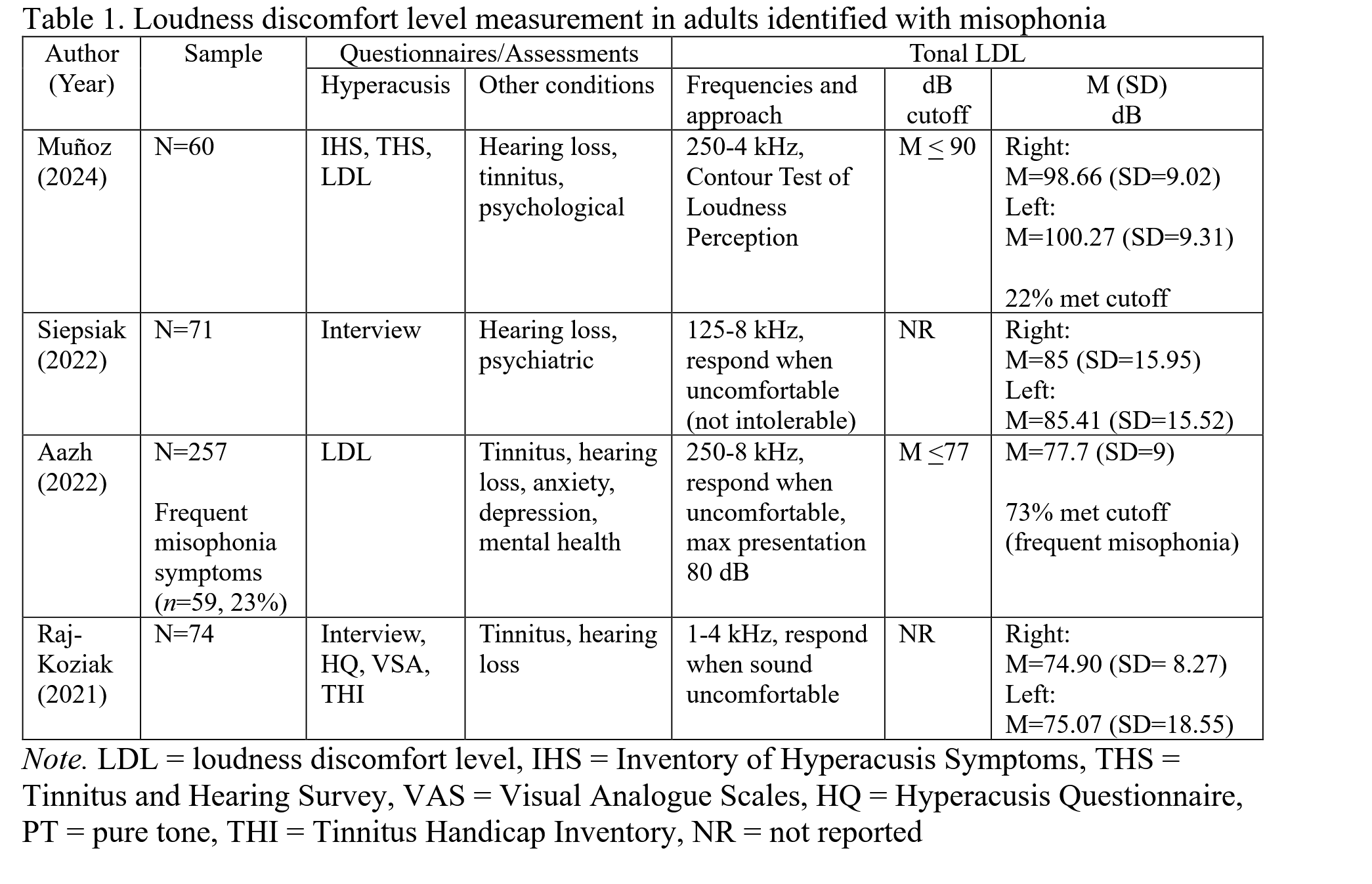
While cautioning against direct comparison of LDLs observed across studies due to no standardized assessments (Woolley et al., 2026b), the Woolley et all discuss how those with co-occurring Misophonia and hyperacusis can have LDLs ranging from 30dB Hl to 120 dB HL, in contrast to the standard < 80dB HL for those with only hyperacusis (Woolley et al., 2026b)
bold-headers
Otalgia predicts hyperacusis severity in children
Keenehan et al. (2026) surveyed parents of 375 pediatric otolaryngology patients aged 3 to 18 years, measuring sound sensitivity with the Hyperacusis Assessment Questionnaire (HAQ) and collecting medical history including reported otalgia (ear pain), then modeled HAQ predictors with multivariate linear regression. They found that frequent otalgia was the strongest predictor of elevated hyperacusis scores, and otalgia also predicted misophonia presence in a separate logistic model. The authors concluded that otalgia may serve as both a symptom and a clinical marker of hyperacusis in children. (Keenehan et al., 2026).
headerless
Body
in a 2026 study, Roy et al found normal ILD thresholds alongside elevated ITD thresholds in Misophonics vs controls, suggesting that intensity-based spatial processing is preserved while temporal binaural processing is affected (Roy et al., 2026)
headerless
Body
Vijayakumar & Ahmmed 2025 implications for audiological traits in Misophonia
A 2025 study evaluating audiometric assessments found that while high-frequency thresholds were lower than low-frequency thresholds across all groups, only the misophonia with hyperacusis group showed a significantly steeper high-to-low slope in the right ear. SRTs did not differ between groups. ULLs were lower in DST groups, especially at 4 kHz, but did not correlate with daily life impact. Tinnitus was more common in DST groups and, in the no DST group, was linked to lower 4 kHz ULLs in the right ear. Past OME history showed no group differences. Overall, routine audiological measures could not reliably differentiate misophonia from hyperacusis. (Vijayakumar & Ahmmed, 2025)
headerless
Body
In a 2024 audiological assessment of 60 adults with misophonia, Muñoz et al. found that 97% had normal hearing thresholds, though 15% showed mild speech-in-noise (SNR) loss, 25% had tonal LDLs suggestive of hyperacusis, and self-reports indicated elevated misophonia severity (MQ mean = 38.05; 58% ≥ moderate), with 12% and 77% reporting clinically significant hyperacusis and hearing problems, respectively. Despite normal audiometric profiles and excellent word recognition, many participants reported difficulties with sound tolerance, speech perception in noise, and tinnitus, highlighting a mismatch between objective audiological data and subjective experience. The authors concluded that misophonia cannot be fully captured through standard hearing tests alone and emphasized the need for standardized, multidimensional assessment protocols that account for perceptual and affective symptoms. (Muñoz et al., 2024)
headerless
Body
Spectral modification cut trigger aversiveness while temporal changes did not, implicating spectral cues averaged over ~250 ms in misophonic triggers.
in a 2024 paper looking at the impact spectral and temporal modification of trigger and neutrals sounds impacts aversiveness, the authors found that spectral modification significantly reduced aversiveness of trigger sounds compared to temporally modified and unmodified sounds. this finding, taken with the lack of effect of temporal modification of 25-ms within 250ms radius on aversiveness, the authors suggest that spectral information averaged over 250ms is important for triggers in Misophonia. (Kazazis et al., 2024)
- Note that I do not fully think I understand this logic
headerless
Surja findings of normal audiological functioning in Misophonia
In a 2023 study, Suraj assessed linear OHC, non-linear OHC, and MOCB efferent functioning in misophonia by comparing TEOAE amplitudes, DPOAE amplitudes, and contralateral suppression magnitudes between misophonic and non-misophonic adults with normal hearing. They found no group differences in absolute OAE amplitudes or in suppression magnitudes for either TEOAEs or DPOAEs at any frequency. From this they concluded that cochlear OHC mechanisms and MOCB efferent functioning are not major contributors to misophonia, and that the pathophysiology likely lies more centrally within the auditory and limbic systems (Suraj et al., 2023).
## proposed -
headerless
Body
in a 2022 study, Aazh notes the theoretical rational behind the idea that hearing loss could impact the Misophonic experience. for instance, they note the tendency of classic Misophonic triggers to be dominated by higher frequency components, and suggests that hearing loss at such frequencies would reduce the audibility of, and thus sensitivity to, such sounds. they contrast this point with a brief mention of loudness recruitment, or a faster perception of gains in loudness for every unit of actual loudness increase beyond hearing threshold. with this context, sounds right above threshold could be disproportionately perceived as annoying, such as in Misophonia (Aazh et al., 2022).
# 6. Regulation & Intervention Mechanisms
7. Learning & Sensitization Processes
headerless
Body
Ward 2022 findings of heighted sensitivity to conditioned cues and enhanced discrimination learning in Misophonia
In their 2022 study, Ward et al. examined whether misophonia symptoms were associated with overgeneralization, sharpened tuning, or all-or-nothing discrimination during auditory aversive conditioning. Behaviorally, the conditioned stimulus (CS+) elicited more negative and arousing ratings during acquisition, and higher MSS scores were linked to greater overall negative and arousing responses, with no evidence of behavioral overgeneralization. EEG results showed robust parieto-occipital alpha-band suppression to tone onset, with greater CS+-specific alpha reduction during acquisition that was best fit by an all-or-nothing discrimination model. Higher MSS scores were associated with stronger CS+-specific alpha suppression, indicating enhanced neural discrimination rather than generalized responding. The authors concluded that misophonia symptoms are not characterized by overgeneralization but instead reflect heightened sensitivity and increased attentional engagement to conditioned threat cues, with neural evidence supporting enhanced discrimination learning. (Ward et al., 2022)
## issues with sensitization/emotional dysregulation -
headerless
Body
In a 2024 TMS study examining neurostimulation and cognitive restructuring for emotional distress in individuals with misophonia and emotional dysregulation, Neacsiu found that misophonic adults reported greater distress and more difficulty downregulating after trigger sounds, whereas clinical controls were more bothered by general aversive sounds. The authors interpreted this as sensitization: when someone is highly tuned to a specific sound category, that sound elicits an exaggerated perception of distress that can exceed the actual physiological response, reflecting a learned or heightened sensitivity rather than a parallel bodily reaction. (Neacsiu et al., 2024)
8. Methodological & Contextual Factors
issues with behavioral index of Misophonia severity
headerless
Body
visual flanker may not be good Misophonia task
in a 2025 study using an oddball paradigm, the authors found lack of behavioral data supporting abnormal attention but did find EEG results supporting it (reduced N1) in line with (Schröder et al., 2014). this mismatch lead them to conclude that either the Behavioral impairments in misophonia are to subtle to detect through behavioral measures, or that the specific measure they used (a visual flanker) is not appropriate to look at the deficiencies in Misophonia specifically. (Patro et al., 2025)
possible impact of anticipatory effects
headerless
Body
it is possible that vigilance and expectations could impact the task based results of studies on Misophonics
in their 2025 literature review, Savard et all note that Misophonics who expect to get triggered when going into study might start with a higher baseline arousal then those who don't,. This could impact the ability of the subjects to focus on cognitive tasks and affect baseline physiological measurements in ways not accounted for by experimental designs (Savard & Coffey, 2025)
9. Genetic & Heritability Findings
bold-headers
Misophonia Symptom Shows SNP Heritability with Loci Near TENM2 and NEGR1
Smit et al. (2023) functionally annotated a genome-wide association study (GWAS) of a single rage-to-chewing-sounds self-report item, used as a misophonia proxy, in 80,607 European-ancestry participants from a general-population 23andMe sample. They identified two genome-wide significant loci, one on chromosome 5 near the TENM2 gene and one on chromosome 1 near an expression quantitative trait locus for NEGR1, alongside substantial single-nucleotide-polymorphism (SNP) heritability, though gene-based and transcriptome-wide analyses did not converge on a consistent risk-gene account. The authors concluded that these annotations, while heritable, did not point clearly to specific risk genes or neural mechanisms (Smit et al., 2023).